During GPCR agonist discovery, receptor desensitisation can significantly affect a compound’s efficacy, particularly following repeated dosing. Understanding how agonists influence receptor signalling and desensitisation is therefore critical when selecting and optimising candidate molecules.
BioAscent scientists used the Agilent xCELLigence platform to monitor real-time cellular impedance changes in HT-29 cells, which endogenously express GPR35b, in response to agonist. A series of pharmacological approaches were applied, enabling comprehensive characterisation of the desensitisation profile of endogenous GPR35b in real-time.
This study demonstrates how label-free impedance technology can provide detailed insights into GPCR signalling that are not accessible from conventional endpoint assays. Using an endogenous expression system allowed physiologically relevant assessment of GPR35b desensitisation, recovery and signalling pathway utilisation, generating a deeper understanding of receptor behaviour to support hit optimisation in agonist discovery programmes.
GPR35 is an orphan G protein-coupled receptor implicated in inflammatory disorders, gastrointestinal disease and cancer, making it an attractive therapeutic target.1-3
While potency and efficacy are important parameters during agonist discovery, these measurements alone may not predict long-term receptor behaviour. As with many GPCR targets, prolonged or repeated agonist exposure can trigger receptor desensitisation through phosphorylation by G protein-coupled receptor kinases (GRKs) and the subsequent recruitment of β-arrestins. Understanding receptor desensitisation is essential when developing agonists intended for chronic administration, as compounds that rapidly desensitise receptors may lose efficacy over time.
The Agilent xCELLigence platform is a powerful label-free approach which monitors GPCR signalling through changes in cellular impedance. A real-time kinetic assay, it is particularly valuable for investigating dynamic processes such as receptor desensitisation and recovery.
In GPCR drug discovery, initial screening campaigns often generate multiple hit series with comparable potency and efficacy in conventional functional assays. However, compounds with similar activity can differ significantly in their signalling kinetics, receptor desensitisation profiles and ability to maintain efficacy following repeated dosing.
These differences may emerge later in development, after substantial medicinal chemistry resources have been invested. Identifying compounds that produce durable pharmacological responses and minimise receptor tolerance to desensitisation earlier in the discovery process can therefore improve candidate selection and reduce development risk.
Although there has been extensive research into the mechanism of desensitisation, when studying a GPCR target it can be challenging to understand because the process comprises many molecular events across different timescales.
BioAscent scientists utilised GPR35b endogenously expressed in HT-29 cells to establish whether real-time cellular impedance could be used to characterise receptor desensitisation in this physiologically relevant endogenous model system. Specifically, the project aimed to:
Determine whether agonist pre-exposure induces GPR35b desensitisation.
Investigate the role of GRK5/6-mediated receptor phosphorylation.
Understand the contribution of β-arrestin-2 to receptor regulation.
Confirm the downstream signalling pathways responsible for impedance responses.
BioAscent scientists utilised HT-29 cells, which endogenously express GPR35b, and monitored impedance responses following stimulation with the known GPR35 agonist lodoxamide.
Agonist-induced receptor desensitisation
Cells were pre-treated with different concentrations of lodoxamide in a concentration-response experiment. Pre-exposure to agonist produced a dose-dependent reduction in both potency (pEC50) and maximal response (Emax), demonstrating that GPR35b undergoes significant desensitisation following activation (Figure 1).
Importantly, the real-time impedance traces showed that desensitised receptors generated smaller, less sustained responses than untreated controls, highlighting the value of monitoring the full signalling profile rather than relying on a single endpoint measurement.
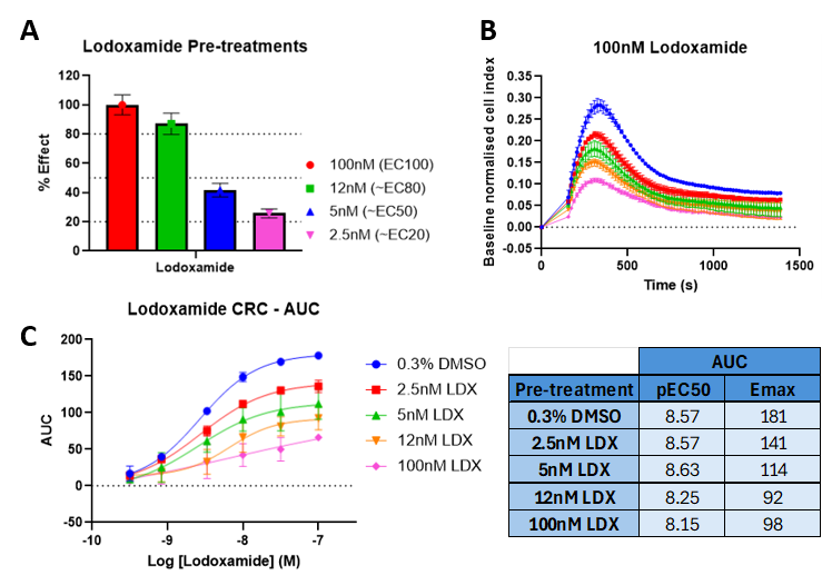
Figure 1: Cellular impedance in HT-29 cells from a CRC to GPR35 agonist lodoxamide after 20 min pre-treatment with different lodoxamide concentrations and 0.3% DMSO wash-out for 30 min: A) actual lodoxamide pre-treatment concentrations as % effect, B) baseline normalised impedance traces for 100nM lodoxamide for each pre-condition concentration, demonstrating the time course data obtained from which area under the curve was calculated in GraphPad Prism, and C) area under the curve values versus lodoxamide concentrations tested for each condition. A 4-parameter non-linear regression curve fit with variable slope was applied where pEC50 and Emax values are reported in the table.
GRK5/6 regulates GPR35b desensitisation
To investigate the mechanisms driving receptor desensitisation, cells were pre-treated with the GRK5/6 inhibitor GRK6-IN-1 before agonist application.
GRK5/6 inhibition produced a dose-dependent enhancement of both peak and sustained impedance responses with a rightward shift in the peak response (Figure 2). These data indicate that GRK5/6-mediated phosphorylation plays a central role in GPR35b desensitisation by promoting recruitment of β-arrestin. In contrast, treatment with the GRK2/3 inhibitor Compound-101 (0.5 µM) had no measurable effect on the impedance response (data not shown), suggesting that GRK2/3 contributes little, if at all, to GPR35b desenstisation in this cellular context.
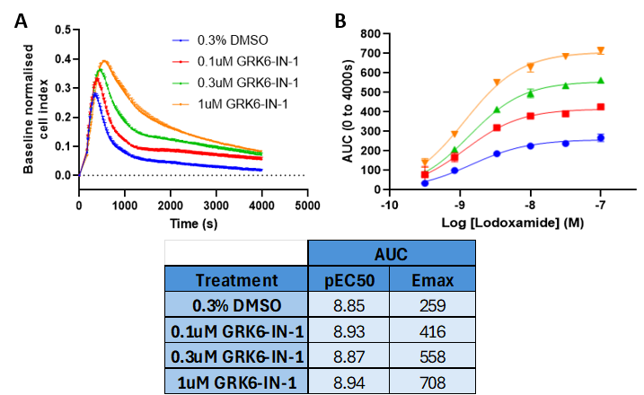
Figure 2: Cellular impedance in HT-29 cells from a CRC to GPR35 agonist lodoxamide after 20 min pre-treatment with different lodoxamide concentrations and 0.3% DMSO wash-out for 30 min: A) actual lodoxamide pre-treatment concentrations as % effect, B) baseline normalised impedance traces for 100nM lodoxamide for each pre-condition concentration, demonstrating the time course data obtained from which area under the curve was calculated in GraphPad Prism, and C) area under the curve values versus lodoxamide concentrations tested for each condition. A 4-parameter non-linear regression curve fit with variable slope was applied where pEC50 and Emax values are reported in the table.
GPR35b desensitisation and recovery
Further experiments combined agonist pre-treatment with GRK inhibition to assess receptor recovery.
Pre-treatment with an EC80 concentration of lodoxamide followed by wash-out substantially reduced subsequent responses to a CRC of lodoxamide, indicating desensitisation of the receptor. The presence of GRK6-IN-1 significantly attenuated this effect, reducing the ability of the agonist to desensitise GPR35b. Recovery of receptor signalling was greatest when GRK6-IN-1 was applied before and after EC80 lodoxamide treatment (Figure 3).
These data demonstrate that GRK5/6-mediated phosphorylation is a major driver of GPR35b desensitisation and that inhibition of this process can partially preserve receptor responsiveness.
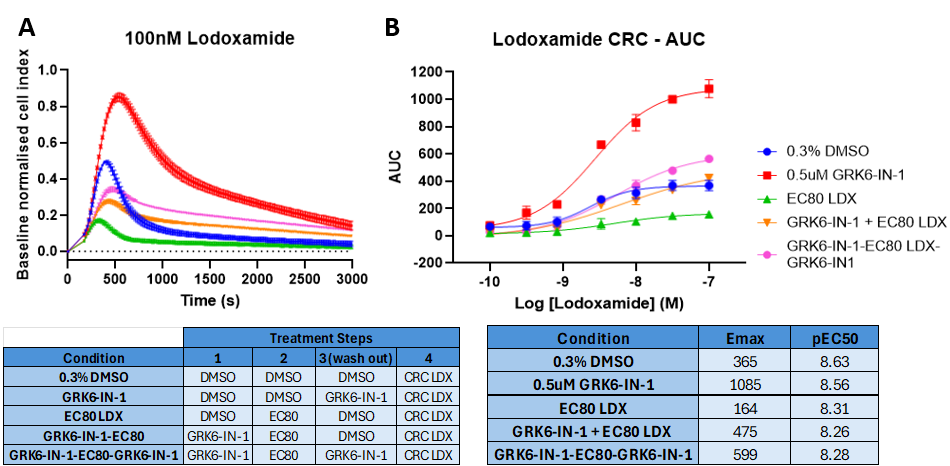
Figure 3: Cellular impedance in HT-29 cells from a CRC to GPR35 agonist lodoxamide after the pre-treatments (30 min for GRK6-IN-1 and 20 min for EC80 lodoxamide) detailed in Table 1, where wash-out was for 30 min: A) baseline normalised impedance traces for 100nM lodoxamide for each pre-condition demonstrating the time course data obtained from which area under the curve was calculated in GraphPad Prism, B) area under the curve values versus lodoxamide concentrations tested for each condition. A 4-parameter non-linear regression curve fit with variable slope was applied and Emax and pEC50 values are shown in Table 2.
β-arrestin-2 acts as a regulatory protein
To further explore receptor regulation, β-arrestin-2 expression was reduced using siRNA-mediated knockdown (Figure 4).
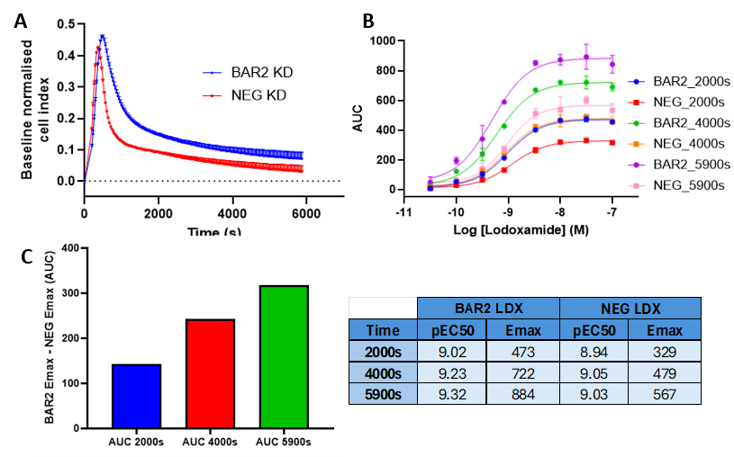
Figure 4: Cellular impedance in HT-29 cells from a CRC to GPR35 agonist lodoxamide 48h after transfection with siRNA for β-arrestin-2 versus a negative scrambled siRNA: A) baseline normalised impedance traces for 100nM lodoxamide for each siRNA transfection, B) area under the curve values versus lodoxamide concentrations for each condition plotted for each time range. A 4-parameter non-linear regression curve fit with variable slope was applied where pEC50 and Emax values are reported in the table. C) The difference in AUC Emax values for BAR2 versus NEG at each of the time ranges.
β-arrestin-2 knock-down increased both peak height and the sustained response without causing a knock-down of signal. Additionally, qPCR analysis on RNA extracted from both the NEG and BAR2 siRNA transfected cells confirmed a 56% knock-down of β-arrestin-2, with consistent levels of GPR35 and GAPDH control in BAR2 compared to NEG cells. These data suggest that β-arrestin-2 is not the primary driver of signalling in this case but instead functions predominantly as a regulatory protein that contributes to receptor desensitisation where its removal allows continued G12/13 signalling.
Confirmation of G12/13-ROCK signalling
Previous reports have suggested that when stimulated by lodoxamide, GPR35 signalling in HT-29 or HEPG2 cells is driven predominantly through G12/13 which signals through the RhoA/ROCK pathway.4,5 To confirm this mechanism, cells were treated with the ROCK1/2 inhibitor Y27632.
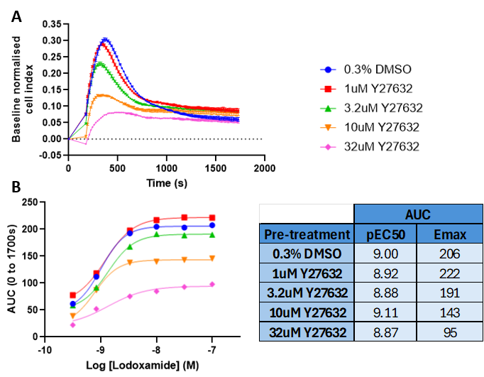
Figure 5: Cellular impedance in HT-29 cells from a CRC to GPR35b agonist lodoxamide after 30 min pre-treatment with different Y27632 concentrations: A) baseline normalised impedance traces for 100nM lodoxamide after each treatment. B) area under the curve values for ~30 min versus lodoxamide concentration. A 4-parameter non-linear regression curve fit with variable slope was applied where pEC50 and Emax are reported in the Table.
ROCK inhibition produced a dose-dependent reduction in impedance responses, with the greatest effect observed on the initial peak response, confirming that G12/13-RhoA-ROCK signalling is the primary pathway responsible for the lodoxamide-induced impedance response in HT-29 cells (Figure 5).
This conclusion was further supported by β-arrestin-2 knockdown studies. ROCK inhibition markedly reduced impedance responses for lodoxamide in both β-arrestin-2 knockdown and negative control (NEG) cells (Figure 6), demonstrating that ROCK signalling remains a key downstream mediator regardless of β-arrestin-2 expression. Furthermore, β-arrestin-2 knockdown increased the overall impedance signal in both DMSO- and lodoxamide-treated cells, consistent with a reduction in receptor desensitisation. Together, these data strengthen the evidence that lodoxamide-stimulated signalling in HT-29 cells is predominantly mediated through G12/13 activation of the ROCK pathway.
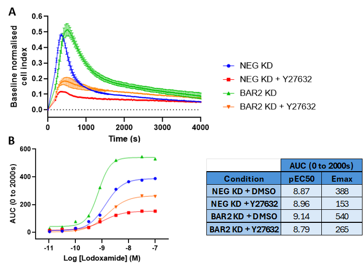
Figure 6: Cellular impedance in HT-29 cells from a CRC to GPR35 agonist lodoxamide 48h after transfection with siRNA for beta-arrestin-2 versus a negative scrambled siRNA. Prior to administration of agonist CRC, cells were pre-treated with 0.3% DMSO control or 32uM Y27632: A) baseline normalised impedance traces for 31.6nM lodoxamide for each siRNA transfection and treatment, B) area under the curve values versus lodoxamide concentrations for each condition. A 4-parameter non-linear regression curve fit with variable slope was applied where pEC50 and Emax values are reported in the table.
This study demonstrates the power of label-free, real-time cellular impedance technology for investigating GPCR receptor regulation in endogenous systems.
Using the xCELLigence platform, our scientists were able to characterise agonist-induced GPR35b desensitisation, identify key regulatory roles for GRK5/6 and β-arrestin-2, and confirm the dominant contribution of G12/13-ROCK signalling in HT-29 cells. The kinetic nature of the assay enabled subtle changes in signalling dynamics to be detected that would likely be missed using conventional endpoint approaches.
For drug discovery programmes targeting GPCRs, these findings highlight the importance of assessing receptor desensitisation alongside initial efficacy measurements. By incorporating physiologically relevant cellular models and real-time functional assays, BioAscent scientists gain a more complete understanding of target biology, differentiate compounds within a hit series, and generate meaningful SAR beyond potency, allowing our clients to make better-informed decisions when selecting agonist candidates for progression.
Learn more about GPCR Drug Discovery at BioAscent here.
The work in this case study was performed by BioAscent Senior Scientist Alison Porter.
With over 20 years’ experience working across Big Pharma, academia, and the CRO sector, Alison is a highly experienced pharmacologist with deep expertise in GPCR biology. She has considerable expertise in developing and running complex cell-based assays to support drug discovery programmes.

1. Quon et. al. Therapeutic Opportunities and Challenges in Targeting the Orphan G Protein-Coupled Receptor GPR35. ACS Pharmacol. Transl. Sci. 2020, 3, 801-812. https://doi.org/10.1021/acsptsci.0c00079
2. Wu et. al. GPR35 acts a dual role and therapeutic target in inflammation. Frontiers in Immunol. 2023. https://doi.org/10.3389/fimmu.2023.1254446
3. A. E. MacKenzie et. al. GPR35 as a Novel Therapeutic Target. Frontiers in Endocrinol. 2011, 2:68. https://doi.org/10.3389/fendo.2011.00068
4. Lin et. al. G Protein-Coupled Receptor GPR35 Suppresses Lipid Accumulation in Hepatocytes. ACS Pharmacol. Transl. Sci. 2021, 4, 1835-1848. https://doi.org/10.1021/acsptsci.1c00224
5. Evans et. al. GPR35 signals through G12/13 and ROCK in HT-29 human colonic epithelial cells. British J. Pharmacol. 2023, 180 (S1) 692-694. https://doi.org/10.1111/bph.16109